30 May 2025
  • Blog
  • About
  • Contact
   
  • Current Topics
  • Social Media
  • Innovation
  • Medical Education
  • Residency
  • Research
Headline News
  • Doctors Day 2020 – Anything But Happy 31 Mar 2020
  • Social Media and Professionalism – Protecting Your Patients, Your Profession, and You 30 Oct 2018
  • Reducing the Disclosure Effect in the Vitreoretinal Fellowship Match 24 Oct 2018
  • The Retina Society – 2018 Meeting Highlights 15 Sep 2018
  • Vitreoretinal Fellowship Match Survey – JAMA Ophthalmology 07 May 2018
  • home
  • blog
Branch Retinal Artery Occlusion

Branch Retinal Artery Occlusion

16 Jul 2017 eyesteve 0 7559

This is the first of a series of “Interesting Cases” posts (and videos) which I hope to publish here on EyeSteve.com. My goal with these videos is to, gradually, develop a library of short, concise videos which describe common (and even not-so-common) eye diseases using real-patient details and images, followed by a brief discussion of the disease and its most important features.

In this post (and video!), I will discuss branch retinal artery occlusions.

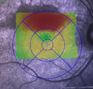
This is a fundus autofluorescence and macular thickness map of a 60 year old African American woman who complained of a black cloud in her inferior visual field which she noticed the prior day. She was hypertensive but had no other vascular risk factors. Her vision was 20/30 at this time. Here you can see hypoautofluorescence and thickening of the superior macula, suggestive of a branch retinal artery occlusion.

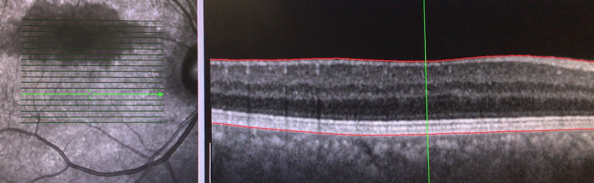
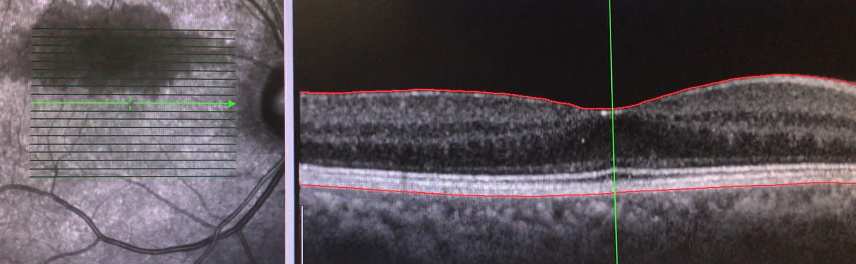
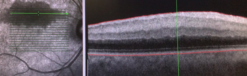
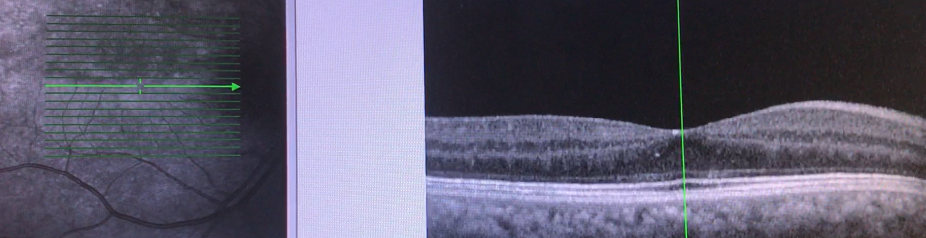
Here are several line scans through the macula. As you can see, the inferior macula is fairly normal, but the inner retinal layers show hyperreflectance and edema of the superior fovea and macula. Remember that the edema is isolated to the inner retina because this area is perfused by the central retinal artery and its branches.

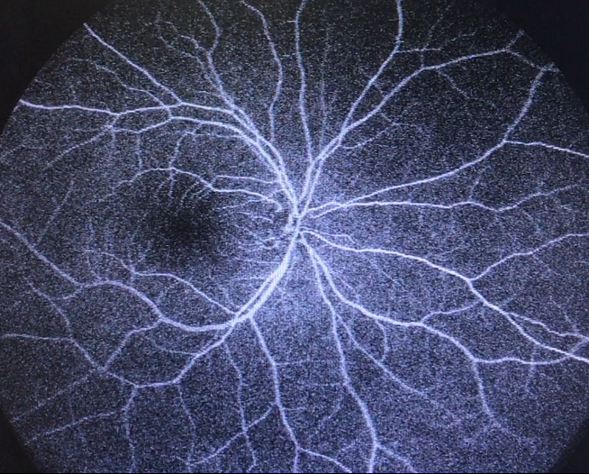
Interestingly, the fluorescein angiogram was essentially normal, without any visible arterial occlusion or area of non-perfusion, possibly because of only a short, transient episode of ischemia, which had already improved by the time she saw us.

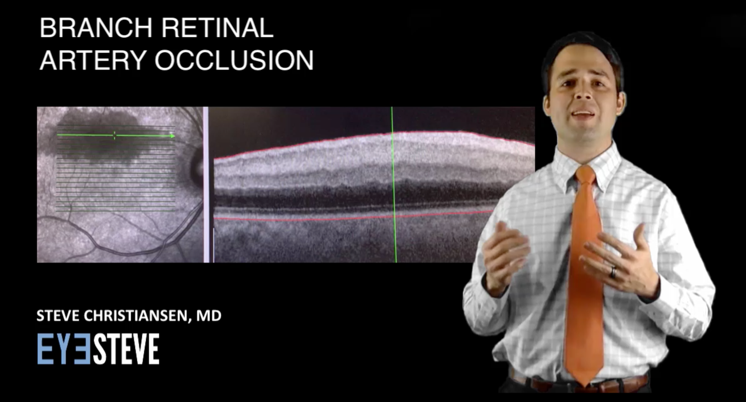
This OCT was taken about a month later, and shows the rapid improvement in the prior hypoautofluorescence and inner retinal thickening.


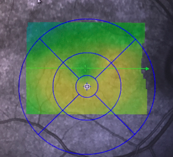
Here is a comparison of the thickness map between the two visits. Vision had improved at this time to 20/25 and she had noticed a lessening of the black cloud in her vision.

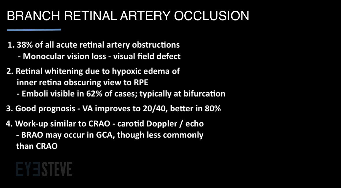
Let’s review just a few pearls of branch retinal artery occlusions. First, it is felt that BRAO’s comprise 38% of all acute retinal artery obstructions. They typically present with monocular vision loss and a relative visual field defect. Secondly, the fundus will have an area of whitening, possibly along the course of a branched arteriole as seen in this fundus photo from EyeRounds.org. Emboli are visible in the majority of cases, typically at vessel bifurcations. Typically BRAOs have a good visual prognosis, with vision improving to 20/40 or better in 80% of cases. The workup for BRAOs is similar to that of CRAOs, with carotid Doppler and cardiac echo to search for embolic sources. Finally, BRAOs can be seen in giant cell arteritis, so be sure to ask about GCA-risk factors, though BRAOs occur less commonly in GCA compared to CRAOs.

Related

OphthalmologyRetina
SHARE THIS ARTICLE :
Tweet

We would love to hear what you think! Post your questions or comments below!Cancel reply

ABOUT THE AUTHOR

eyesteve
  • ABOUT ME
  • OTHER ARTICLES
Steve Christiansen, MD, is a vitreoretinal surgeon practicing in Colorado Springs, Colorado. He previously completed ophthalmology residency at the University of Iowa Department of Ophthalmology & Visual Sciences in Iowa City, Iowa and fellowship at the Cincinnati Eye Institute in Cincinnati, Ohio.

Doctors Day 2020 – Anything But Happy

31 Mar 2020 eyesteve 1

Social Media and Professionalism – Protecting Your Patients, Your Profession, and You

30 Oct 2018 eyesteve 0

Reducing the Disclosure Effect in the Vitreoretinal Fellowship Match

24 Oct 2018 eyesteve 0

The Retina Society – 2018 Meeting Highlights

15 Sep 2018 eyesteve 2
PREVIOUS

The Truth Behind the 27 Contact Lenses Stuck in the Patient's Eye

NEXT

Six tips to land your first (or next!) job - from an ophthalmology recruiter!

RELATED POST

Doctors Day 2020 – Anything But Happy

31 Mar 2020 eyesteve 1

Social Media and Professionalism – Protecting Your Patients, Your Profession, and You

30 Oct 2018 eyesteve 0

Reducing the Disclosure Effect in the Vitreoretinal Fellowship Match

24 Oct 2018 eyesteve 0

The Retina Society – 2018 Meeting Highlights

15 Sep 2018 eyesteve 2

FIND EYESTEVE ON SOCIAL MEDIA

  • Facebook
  • LinkedIn
  • RSS
  • Twitter
  • YouTube

Subscribe to EyeSteve

Latest Posts

Doctors Day 2020 – Anything But Happy

31 Mar 2020 eyesteve 11323

Social Media and Professionalism – Protecting Your Patients, Your Profession, and You

30 Oct 2018 eyesteve 8721

Reducing the Disclosure Effect in the Vitreoretinal Fellowship Match

24 Oct 2018 eyesteve 8252

Latest Tweets From EyeSteve MD

Follow @eyesteve

EyeSteve.com Logo

EyeSteve.com was created to educate, inform, and inspire all things eye by discussing various aspects of medical education, informing patients about eye diseases and innovative new treatments, and inspiring us all toward vision health and appreciation of the wonderful gift of sight.

Recent Posts

  • Doctors Day 2020 – Anything But Happy
  • Social Media and Professionalism – Protecting Your Patients, Your Profession, and You
  • Reducing the Disclosure Effect in the Vitreoretinal Fellowship Match
  • The Retina Society – 2018 Meeting Highlights
  • Vitreoretinal Fellowship Match Survey – JAMA Ophthalmology

Get EyeSteve In Your Email

Subscribe to eyesteve.com and receive notifications of new posts instantly by email.

Follow @eyesteve
View Steven M. Christiansen's profile on LinkedIn
Copyright 2014 | eyesteve.com | All Rights Reserved | Powered by WordPress